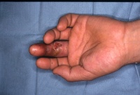
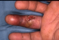
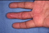
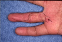
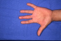
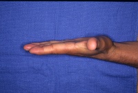
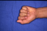
| High
pressure injection injuries of paint, sand, lubricating fluid and
other
materials are uncommon, but important because they are also on the list
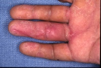
of injuries missed in the accident ward. Typically, the patient has
briefly
placed their hand or fingertip over a pressure spray nozzle, sustaining
an injection of material into the soft tissues. Under pressure, this
material
tracks up tissue planes next to flexor tendons, nerves, arteries and
through
the named bursae and compartments of the hand and arm. Debris may be
driven
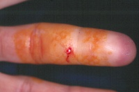
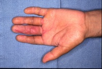
from the fingertip to the chest wall. The examiner may be misled by a
small
visible wound and (depending on the material injected) relatively few
physical
findings, and the patient may be discharged only to return within 24
hours
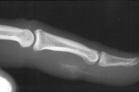
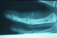
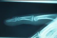
because of worsening symptoms. X-rays may show soft tissue air,
particulate
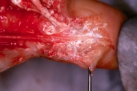
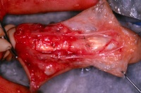
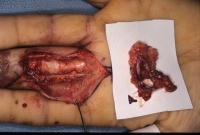
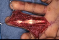
debris, or pigment in certain types of paint. One treatment is
emergency radical
debridement. The pressure injected
material tends to track through the loose areolar tissue along
longitudinal
structures, and only careful debridement may allow preservation of all
vital structures. In
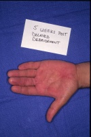
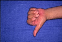
contrast, late surgical treatment may require en bloc tumor
like
excision of contaminated zones or amputation. Late results are worst
when
the injected material is either a petroleum based solvent or
particulate
(sandblasting) material, when the tendon sheath is involved, and when
there
is wide proximal spread of the injected material.
The injected material is not sterile, and prophylactic antibiotic
treatment
is indicated. Pressure injection injuries presenting with poor
perfusion
may be treated with primary amputation.
Injection of pressurized aerosol flurocarbon liquids such as used in
refrigerants
may additionally result in deep frostbite injury. |
