Clinical Example: Osteochondromas of the Distal Phalanx
| Osteochondromas are the most common benign bone tumors. They are believed to arise as a local perichondrial growth abnormality. They usually arise within the first two decades of life and stop growing with skeletal maturity. |
| Click on each image for a larger picture |
| Case 1. This young woman presented with a painless distal phalanx and nailbed deformity which developed slowly during her teens |
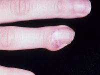
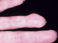
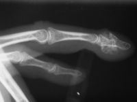
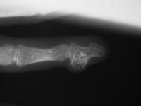
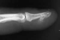
| Plain Xrays show continuity of abnormal bone the with the medullary bone of the distal phalanx. |
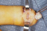
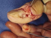
| The tumor was accessed through an eponychial splitting lateral incision. |
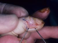
| Te dorsal subungual bone was recontoured. |
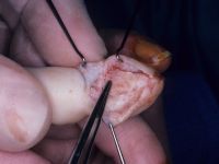
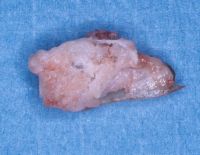
| Specimen. |
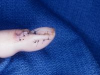
| Eponychial fold reconstruction. |
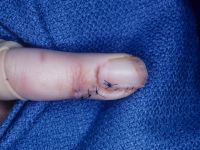
| Case 2. Osteochondroma presenting as a pulp mass. The patient believed that they had a splinter or foreign body in the finger from a forgotten incident. |
| Excision through a midline longitudinal incision.l |
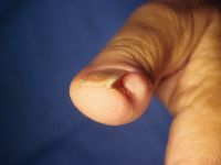
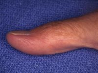
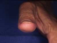
| Case 3. Mass and progressive nail plate contour deformity developed over the last several years in this 42 year old woman. |
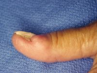
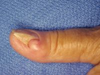
| The nail plate is actually folded against itself in an acute angle. |
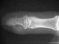
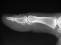
| On plain radiographs, The mass has a central ossification. The lateral border of the distal phalanx has remodelled with traction exostoses at the attachments of the intraosseous ligament. |
| The tumor shelled out with a small proximal attachment. Pathology was interpreted as Nora's lesion: bizarre parosteal osteochondromatous proliferation (BPOP), which is histologically distinct variant of osteochondroma. BPOP has a later age of onset , more rapid growth and more variability than simple osteochondroma. |
| Late result, with normalization of the nail plate contour. |

|
Search
for...
osteochondroma bizarre parosteal osteochondromatous proliferation |
Case Examples Index Page | e-Hand Home |