| Rarely, periarticular
bone growth involves the interphalangeal joints of the fingers. These
cases demonstrate some of the variations. |
| Click on each image for a larger picture |
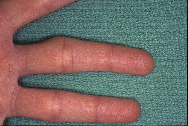
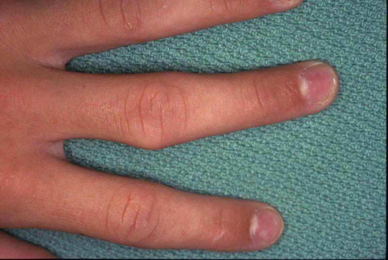
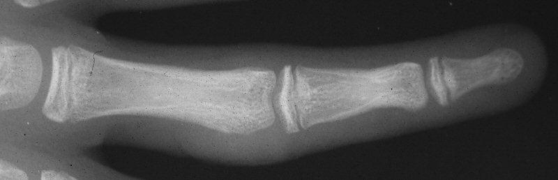
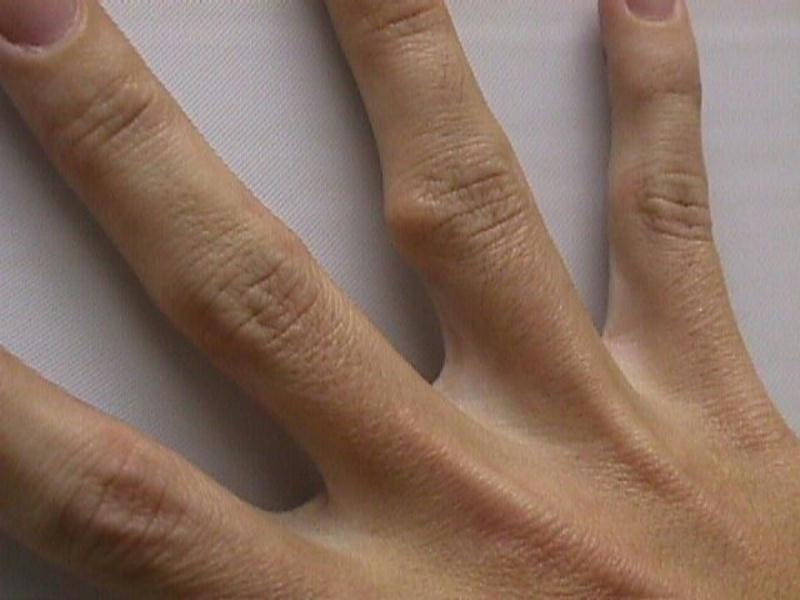
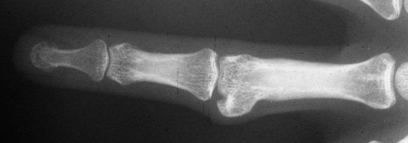
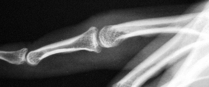
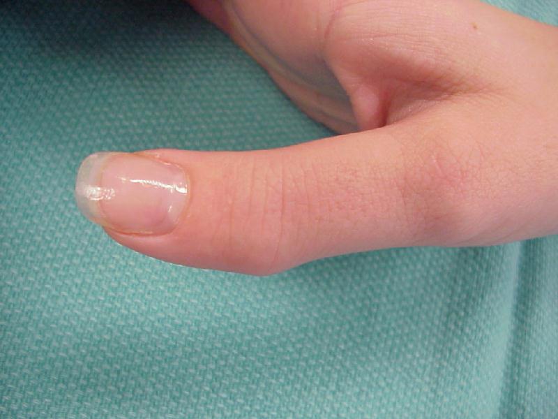
| Case 1. 14 year old boy with radial prominence and ulnar deviation of the middle finger proximal interphalangeal joint. Painless, no history of trauma. |
 |
 |
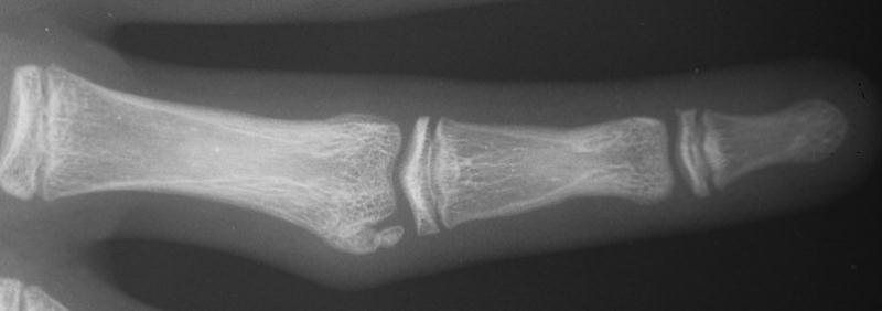
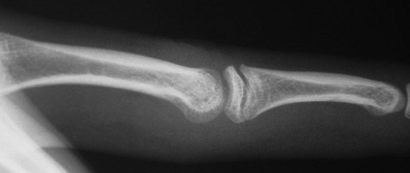
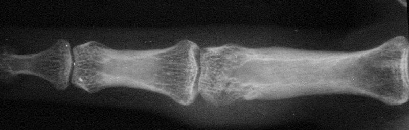
| Radiographs showing well circumscribed calcification at the proximal phalanx collateral ligament origin, 10 degrees of lateral angulation. |
 |
 |
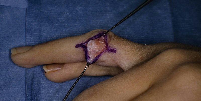
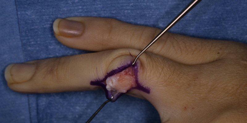
| This was treated with simple excision. Pathology was consistent with mature bone. |
 |
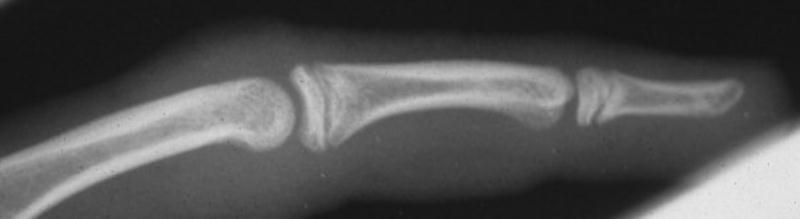 |
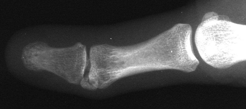
| Case 2. Mass developing after a lateral dislocation of the proximal interphalangeal joint of a 34 year old woman. |
 |
 |
 |
| Radiographs were consistent with either a united collateral ligament avulsion fracture or ossiification of a parosteal hematoma. |
 |
| This was treated with simple excision and collateral ligament repair to local tissues. |
 |
 |
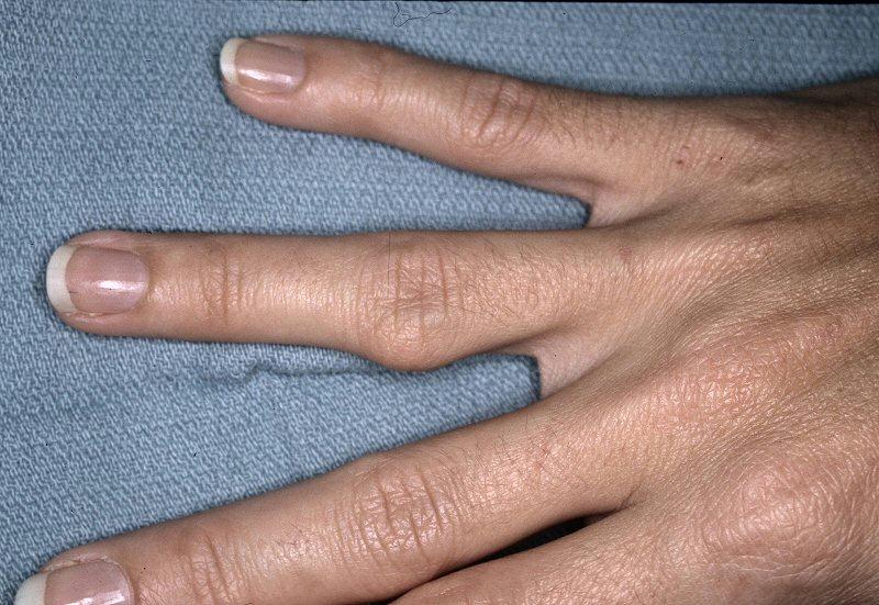
| Late result. |
 |
 |
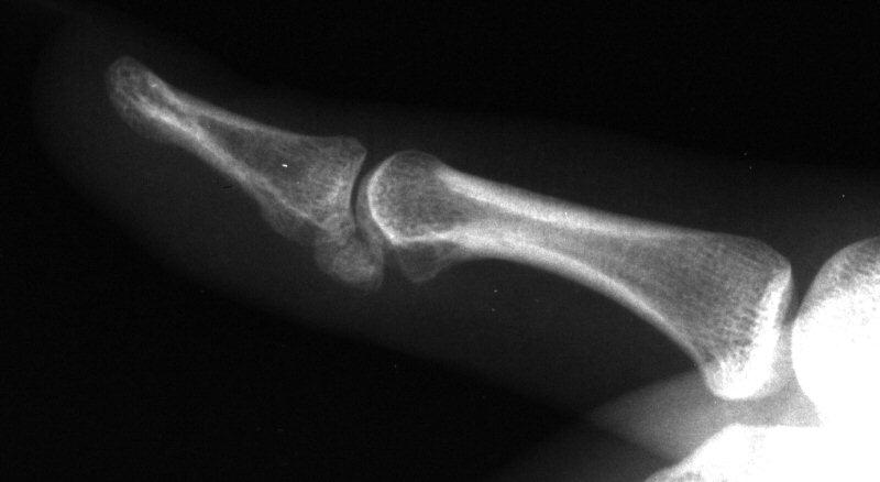
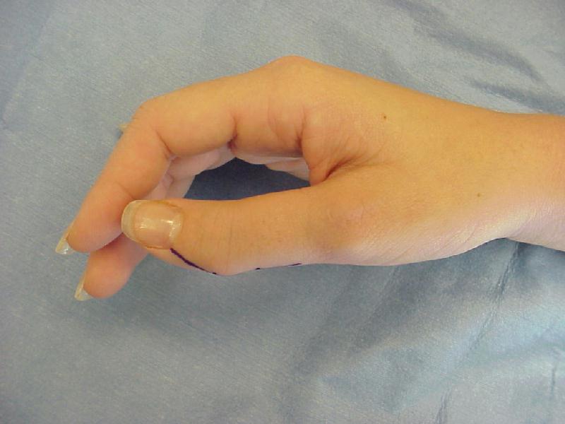
| Case 3. 21 year old woman with pain developing in a congenitally angulated thumb. |
 |
| Radiographs show a juxtaarticular ossification with subchondral cyst formation of the bone interface with the lateral phalangeal head and lateral angulation of the proximal phalanx articular surface. |
 |
 |
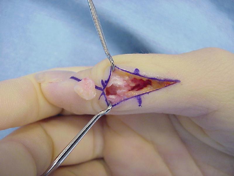
| This was treated with excision of the mass and corrective closing wedge osteotomy of the proximal phalanx. |
 |
| There was no articular cartilage on the pseudojoint, with arthritic type reactive changes. |
 |
| Corrected alignment |
 |
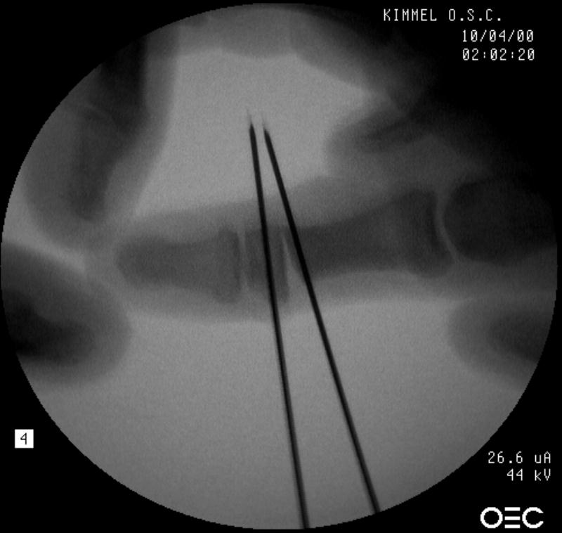
| Intraoperative fluoroscopy. The mass: |
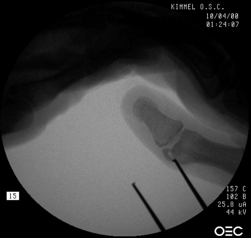 |
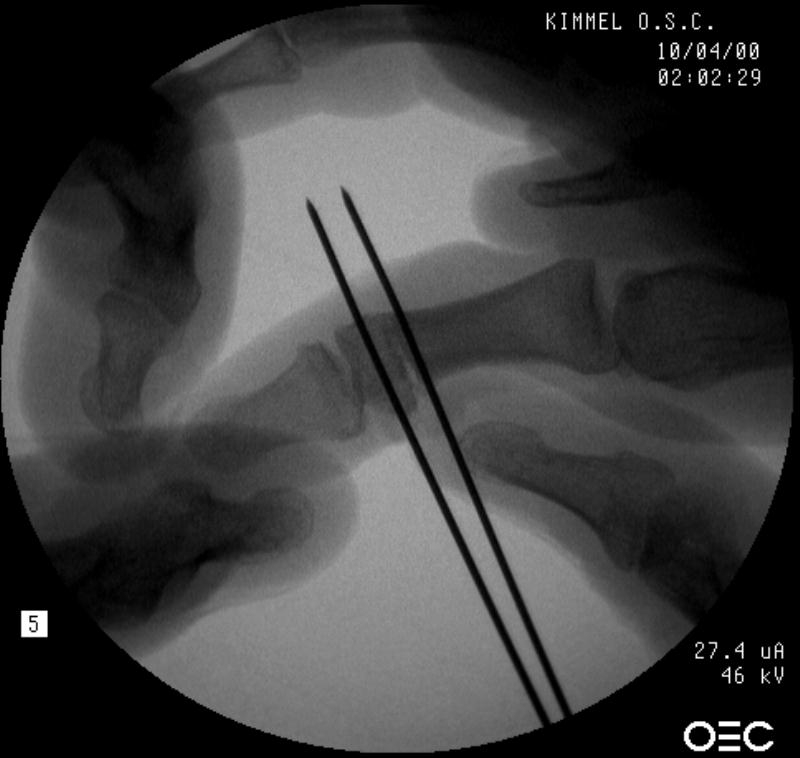
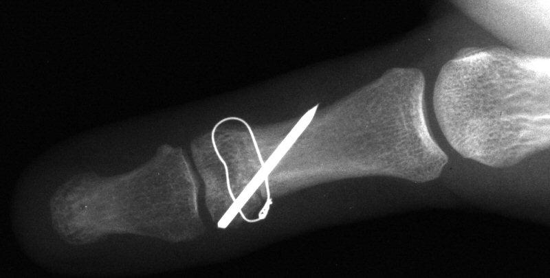
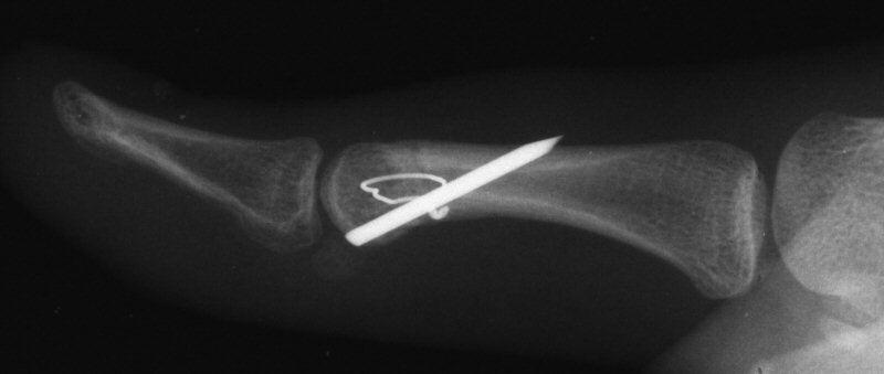
| Osteotomy planning: proximal pin parallel to the proximal joint line, distal pin parallel to the distal joint line: |
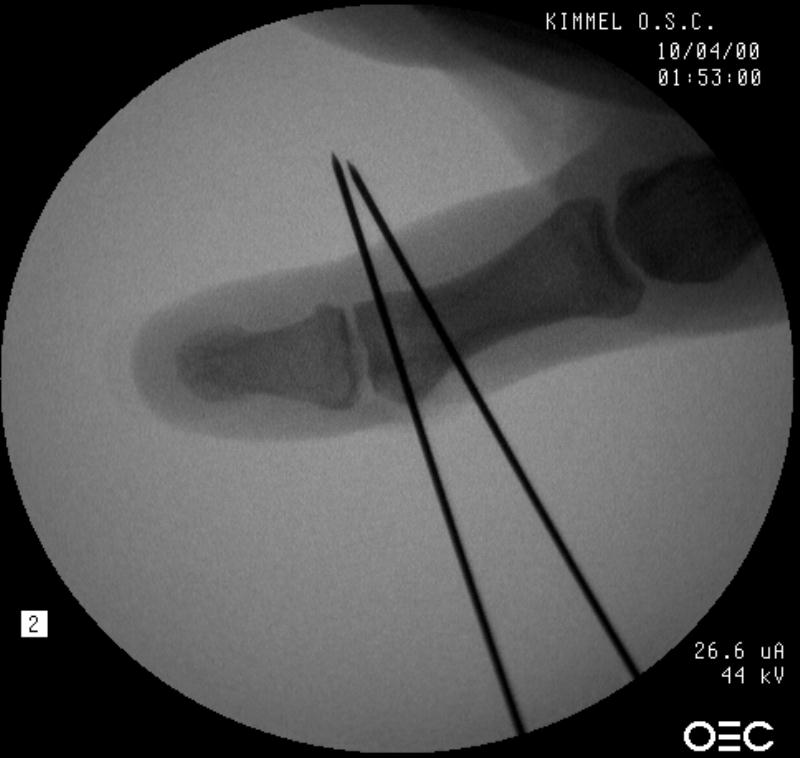 |
| Pins were used as saw blade alignment guides: |
 |
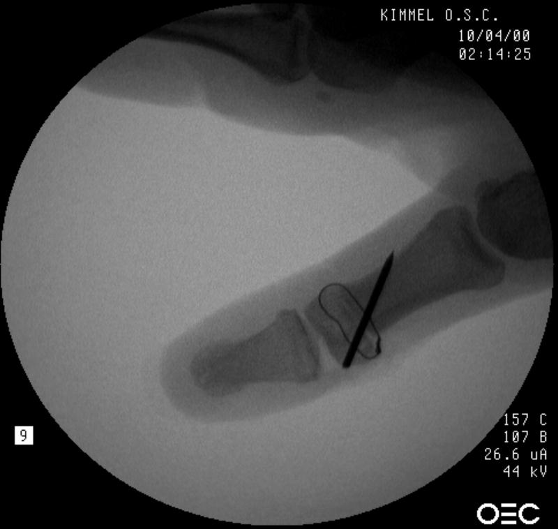
| Osteotomy closed: |
 |
| Intraosseous wire passed through pin tracts, interfragmentary pin: |
 |
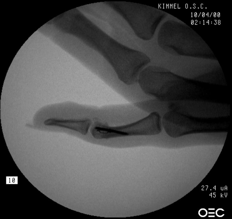 |
| Late result: |
 |
 |
|
Search
for... articular phalanx exostosis phalanx osteotomy |
Case Examples Index Page | e-Hand Home |