Clinical Example: Bilateral complex syndactyly reconstructed in three stages
| Every congenitally different hand is ...different, and requires a customized approach. The reconstruction of complex complete multiple digit level syndactyly was performed in stages as three bilateral operations and illustrates some technical points applicable to other situations. |
| Click on each image for a larger picture |
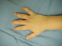
| Bilateral middle, ring and
small acrosyndactyly. |
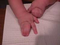
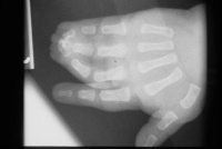
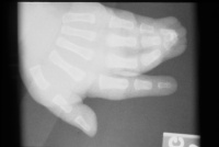
| All procedures were
bilateral at the same setting, but are illustrated here only on the
right side. |
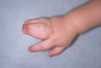


| First stage: fingertip
separation and surfacing with hypothenar skin grafts. This converts the
syndactyly from complex to simple and buys some time for growth by
lessening the deforming forces of the terminal bony syndactyly. |
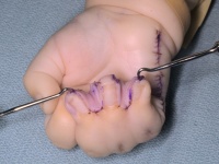
| Postop result. |
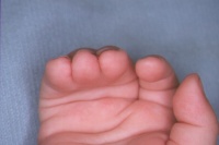
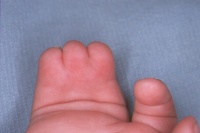
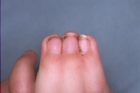
| Angular tethering has been
lessened. |
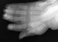
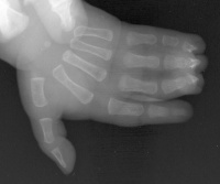
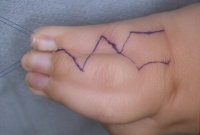
| The second stage was
syndactyly release of the two digits with the greatest length
discrepancy (ring and small) to reduce further angular growth
disturbance from tethering. |
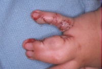
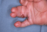
| After healing: |
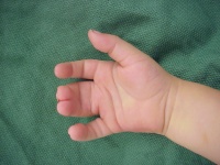
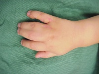
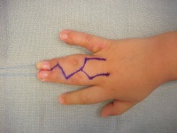
| The third stage: separation
of the remaining digits. |
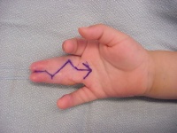
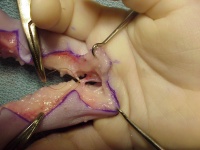
| Neurovascular anomalies,
such as distal common digital bifurcation, are more common in complex
than simple syndactyly. In this situation, the nerve facicles may be
separated by dissection, but the digital arteries can not. One
option is to divide the common digital artery to the digit which has an
undisturbed contralateral vessel, as shown here, dividing the ulnar
digital artery to the middle finger to preserve circulation to the
ring, which has been dissected on both sides. |
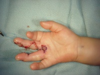
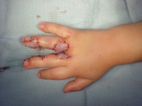
| Postoperative result. |


|
Search for... complex syndactyly acrosyndactyly staged fingertip separation |
Case Examples Index Page | e-Hand home |